Post-acute sequelae of COVID-19 (PASC) is common, but clinicians have found these sequelae, like the acute disease, to be a moving target. Now, however, following two years of experience and mounting radiologic studies, clinicians are narrowing in on some prognostications.
Carla M. Sevin, M.D., director of the ICU Recovery Center at Vanderbilt University Medical Center, works on the forefront of the search for patterns in PASC, dovetailing her current research with a career-long focus on recovery from critical illness.
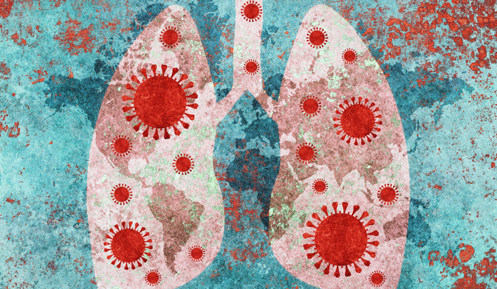
In spring 2022, she gave a talk to the Society of Thoracic Radiology about post-COVID radiological findings, pulmonary function tests, and clinical lung problems in patients with persistent problems after COVID-19. She noted that although some similarities exist between the pulmonary sequelae of COVID-19 and other respiratory infectious diseases, several patterns have emerged that deviate from those commonly observed with other respiratory viruses, such as the flu. Even in mild COVID-19, the lungs are disproportionately vulnerable to damage.
“If you took everybody who was hospitalized with COVID and saw them three months out, perhaps 75 percent of them would have an abnormal chest CT,” she said. “Even among people who were not sick enough to be hospitalized, about 20 percent would have an abnormal CT.”
Yet Sevin says patient responses to COVID-19 and their recovery trajectories are still highly variable.
Predictable Ravages
Lung imaging in recovering patients may show expected changes or reveal dangerous new conditions that need to be monitored. Chief among these, Sevin said, is persistent and recurrent clotting, one of the top risks for patients who have or are recovering from COVID-19.
Post-COVID imaging is also elucidating structural changes such as tracheal damage in those who underwent intubation and even in those who did not, suggesting an effect of the virus on the airways.
“COVID seems to affect the tracheal wall directly and indirectly, so we are seeing a lot of floppy airways and tracheal narrowing and scarring, and these are hard to heal,” Sevin said.
Red Herrings
The discord between symptoms and imaging or test results was unexpected, Sevin said, pointing to the somewhat frequent phenomenon of patients presenting with a lingering shortness of breath or exercise intolerance but having relatively normal pulmonary function and other medical results.
“If you took everybody who was hospitalized with COVID and saw them three months out, perhaps 75 percent of them would have an abnormal chest CT.”
Others can seem to be on the path to recovery when they take a turn for the worse.
“The patient starts off hypoxic, they’re hospitalized and treated, and then they start getting better. Their oxygen requirement is coming down, their CT scans look better,” Sevin said. “But then things start to decline. Sometimes this is a new infection, but can also be organizing pneumonia, a noninfectious process treated with steroids.”
Patients whose COVID-19 landed them in the ICU also are prone to post-intensive care syndrome (PICS), which is often debilitating. These patients, too, can have normal imaging. Younger athletes also may have normal echocardiograms and CTs, even in the presence of COVID-related chest pain.
Medical Miracles
Recovery after COVID-19 has also produced a few “medical miracles,” said Sevin, reflecting on patients with critical cases who temporarily developed severe scarring lung conditions that are typically chronic.
“Before COVID, when we saw pulmonary fibrosis or a scarring pattern on imaging, we did not expect it to go away,” she said. “But we have post-COVID patients with fibrotic scars and cysts on imaging that subsequently get better. It might not completely resolve, but I’ve seen marked reductions in scarring in some patients, and huge cysts that were just like holes in the lung heal up.”
In fact, she says, several patients who were referred for lung transplants were later able to recover without the operation.
Even initial predictions that asthma patients would be at higher risk and have more severe disease were hasty.
“Studies have shown that if somebody in a household gets COVID and their family member has allergic asthma, that family member is not more likely to get COVID. And if they have a food allergy, they’re actually less likely to get COVID.”
Avoiding False Assumptions
Sevin worries that with so many new lung morbidities tied to COVID, many patients assume that any respiratory symptoms they experience after acute infection are complications of COVID and don’t come in for diagnostic imaging and labs.
“I’ve had a number of patients come to me with what they thought was long COVID, but it turned out to be cancer, ALS, or other problems that needed to be diagnosed,” she said.