As low-dose CT lung cancer screening has expanded its reach over the past several years, the costly gulf between nodules with “low and high probability of malignancy” or “low and high probability nodules” has handed the oncological radiology community a clear challenge: how to assess the flood of indeterminate nodules uncovered through advanced imaging and broader screening practices.
This mission has a high profile at Vanderbilt University Medical Center due to a comprehensive, end-to-end lung cancer program that starts with community smoking cessation initiatives and follows through to palliative care. At the leading edge of this effort is research into artificial intelligence (AI) capabilities for creating reliable and clinically pragmatic tools.
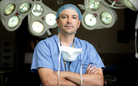
Fabien Maldonado, M.D., a professor of medicine and thoracic surgery at Vanderbilt, is a national leader in AI applications for lung nodule discrimination.
“We now own a deep-learning decision-support tool, Optellum,” he said. “Last month, we diagnosed our first lung cancer patient using its support. We are excited to have this radiomics software that will boost physicians’ confidence in assessing the large gray area of indeterminate nodules.”
Advantage: Deep Learning
Conventional machine learning works through a hand-selected number of radiomic variables believed to be associated with malignancy, Maldonado said. These programs improve classification of indeterminate nodules compared with unassisted readings.
“Through its probability score assignments, Optellum promises to help patch the gap in the way we capture these nodules.”
Deep learning tops this capability. By training on tens of thousands of images with variable characteristics, the algorithm learns to characterize nodules with increasing accuracy. Put to the test, these platforms demonstrated the capacity to reclassify indeterminate lung nodules into low- or high-risk categories in more than a third of cases involving cancers and benign nodules, based on conventional risk model data. Maldonado noted, however, that the field is young, and lung nodule applications will be more efficacious as access to curated datasets grows.
Patching the Risk Gap
Maldonado estimates 20 million chest CT scans are performed annually in the United States. Low-dose CTs decrease the relative risk of cancer-specific mortality among high-risk individuals by 20 percent . High demand for the scans has meant radiologists must work at a pace of three to four minutes per study, Maldonado said. The challenge has become how to capture all the data being collected and processed by the imaging software for the benefit of patients.
Radiomics was born in response to that mission. Its role in early detection can help radiologists assign a larger proportion of indeterminate nodules to “watchful waiting” or “biopsy,” reducing the number that are acted on out of an ill-defined abundance of caution.
“Lung nodules are ubiquitous, and only a very small minority are malignant,” Maldonado said. “PET scans are expensive. Biopsies are invasive. Through its probability score assignments, Optellum promises to help patch the gap in the way we capture these nodules.”
Indolent or Aggressive?
Maldonado and his Vanderbilt colleagues are also working on an NIH grant to study a second radiomic application that would determine, non-invasively, which adenocarcinomas are aggressive versus indolent.
“Adenocarcinoma is the most common form of lung cancer, and while some of these lesions are very aggressive, those that are indolent are often over-diagnosed,” Maldonado said. “Like the case with many prostate and thyroid cancers, the patient may die with the cancer, not from it.”
“Since we do more bronchoscopic lung nodule biopsies than anyone else in the U.S., Optellum was a logical investment for us.”
He says that radiomics-supported characterization of these lesions may enable a more tailored approach to care management. “If the cancer is not going to kill the patient, it is really kind of an incidental finding. On the other hand, if it is a very aggressive one that’s likely to recur in spite of optimal surgical treatment, maybe you need to add adjuvant therapy to that.”
Heading Mainstream
Maldonado first immersed himself in machine learning at Mayo Clinic while working in risk stratification of adenocarcinoma. Through a collaboration with Vanderbilt, he met and later worked in the labs of the late Pierre Massion, M.D., who was director of the Cancer Early Detection and Prevention Initiative at Vanderbilt-Ingram Cancer Center, and former Mayo mentor Otis Rickman, D.O., now director of interventional pulmonology and the Lung Nodule Clinic at Vanderbilt.
“We’re applying the first AI decision-support system in Tennessee, and are one of only two installations in the U.S.,” Rickman said. “The Vanderbilt Lung Nodule Clinic is the only place in the U.S. that is using it clinically,” Rickman said. “Since we do more bronchoscopic lung nodule biopsies than anyone else in the U.S., Optellum was a logical investment for us.”
Maldonado and Rickman say the goal is to extend machine learning to benefit populations well beyond Vanderbilt and the region.
“Ultimately, what we’re trying to do with machine learning is bring everybody to a level playing field, so that even people who don’t have lots of expertise and experience dealing with these nodules will have enough information to make the right decisions,” Maldonado said.
At the Bench
The team’s research continues to push radiomics forward. They are working under NIH R01 funding to evaluate how Optellum probability scores change over time on serial CTs.
In collaboration with Mayo Clinic, they have developed radiomic tools to distinguish benign from malignant nodules, as well as indolent from aggressive lung adenocarcinomas.
“The momentum is huge in our division. We are currently applying for additional NIH funding to further improve the risk classification of lung adenocarcinomas,” Maldonado said.


