Through a major undertaking at Vanderbilt University Medical Center, a team of researchers has collected and catalogued complex images of the human pancreas, creating a scalable, publicly available database for the broader scientific community.
The database, dubbed “PancreatlasTM,” is the world’s first online imaging database of human pancreatic tissue. It includes detailed, multi-layered images linked to deidentified clinical data from children and adults, addressing a long-unmet need of researchers studying an organ that cannot be safely biopsied.
“Pancreatlas is a user-friendly platform where researchers can interact with images, browse or navigate through them, annotate them and bookmark them,” said Marcela Brissova, Ph.D., a research professor of medicine at Vanderbilt and co-scientific director of the project.
Broader Access to Sample Images
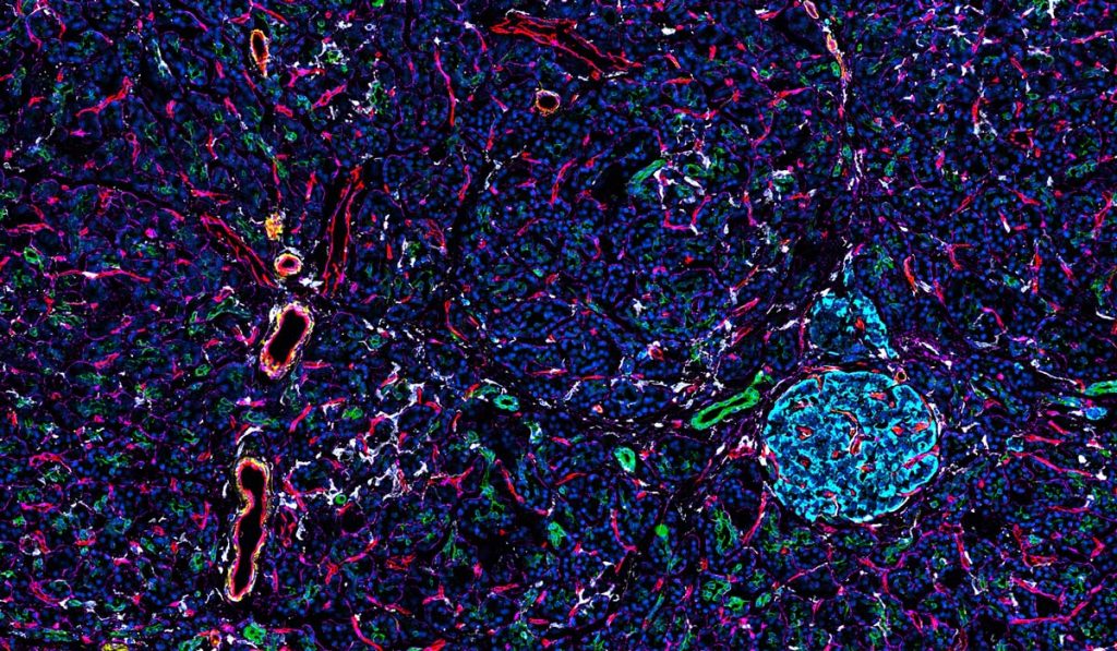
A primary goal of Pancreatlas is to provide a platform to share images collected from cadaveric human pancreas samples. These precious samples are typically sectioned to create a few small, static figures for publication, while additional valuable data they contain go unpublished.
“One of the biggest challenges in the imaging world is that there are not very good tools to manage large imaging datasets.”
Linking images to support broader analyses has been an emerging theme for researchers across specialties. Said Brissova, “One of the biggest challenges in the imaging world is that there are not very good tools to manage large imaging datasets where individual researchers can actually go and interact with images live.”
Brand New Infrastructure
The Pancreatlas team developed a web platform and user interface from the ground up to support the project. Leading the IT efforts is Jean-Philippe “J.P.” Cartailler, Ph.D., director of Creative Data Solutions, a Vanderbilt-shared resource.
“Scientists need cost-effective yet fully featured database solutions that facilitate large dataset sharing in a structured and easily digestible manner,” wrote Cartailler and the team when describing their front-end methodologies in Patterns, a Cell Press publication.
New IT infrastructure enables Pancreatlas to integrate images from Vanderbilt investigators, as well as from the NIH-funded Human Pancreas Analysis Program and the Network for Pancreatic Organ Donors with Diabetes. Three years of design and development later, it now houses over 800 high-resolution images and counting.
“Our filtering menus allow users to view and refine images based on multiple variables of interest.”
A Research Goldmine
Pancreatlas is practically arranged by “Image Collections” based upon common research themes, such as type 1 diabetes or cystic fibrosis-related diabetes. Pancreatlas also contains comprehensive image datasets from across the human lifespan to provide context for investigators working, for example, to understand diabetes’ progression or the origins of pancreatic cancer.
“By housing images in Image Collections, we provide curated points of entry to large image datasets; our filtering menus allow users to view and refine images based on multiple variables of interest,” the team explains in the “About” page on pancreatlas.org.
Site visitors also benefit from detailed metadata associated with each image, which helps to standardize nomenclature and ease searches. Dozens of tags cover disease status, biomarkers, sample collection and processing, imaging modality, and pancreas region, among other key details.
The developers plan to expand the collections of curated images over time and hope to connect additional data, such as genomics and physiological information.
Team Science
In addition to the Vanderbilt Diabetes Center and the Vanderbilt Diabetes Research and Training Center, Pancreatlas partners include other academic institutions and multiple funding agencies.
Diane Saunders, Ph.D., co-scientific director of Pancreatlas, is also quick to mention the most central contributors to the project – donors and families.
“If an organ doesn’t meet the criteria for clinical use such as transplantation, that’s often when we are able to study the pancreas,” she said. “We’re very grateful, especially to the families of these organ donors who must make difficult decisions. We appreciate that these tissues are incredibly valuable resources, and our research community is able to make important discoveries by studying them and sharing them with scientists from around the world via Pancreatlas.”