Virtual reality (VR) is the hot technology for surgical planning and training, but it is still a newcomer in pediatric cardiac surgery. At Monroe Carell Jr. Children’s Hospital at Vanderbilt, the congenital heart disease (CHD) team is piloting VR tools for surgical planning thanks to the talents and generosity of one patient’s father.
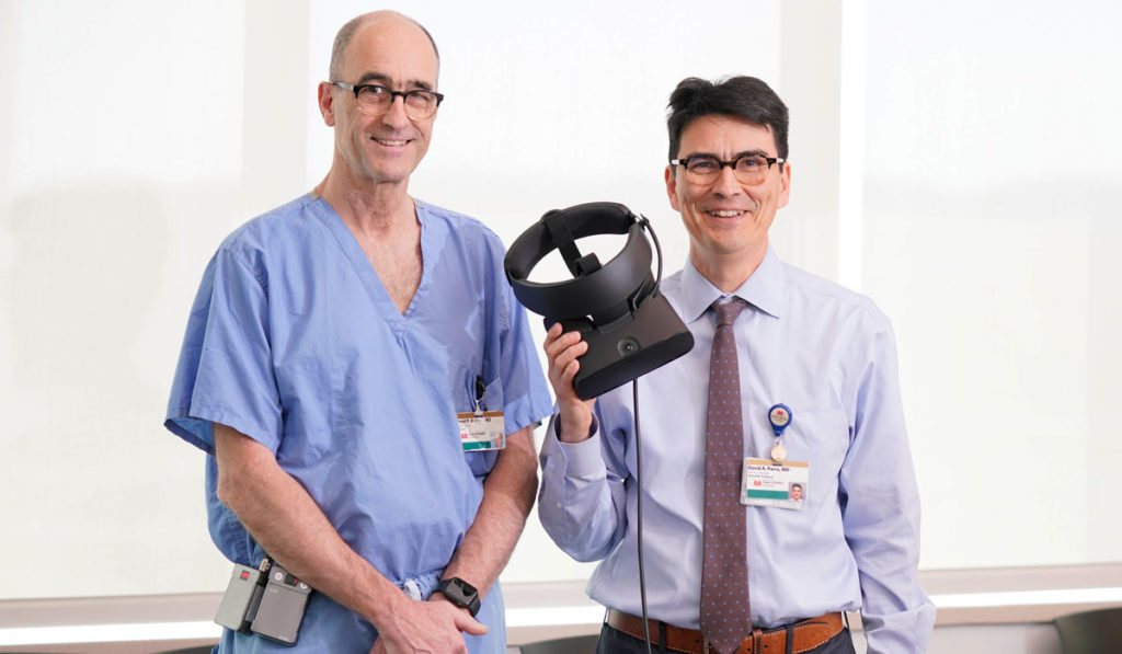
After their son Calvin was born with a single ventricle, Tyler Thayer and his wife Erica started Project Heart, a foundation that funds CHD research grants. When Calvin underwent a Fontan procedure at Children’s Hospital, Thayer, an architect, and David Parra, M.D., director of cardiac imaging, discovered a “lock and key” opportunity: linking Parra’s interest in VR with Thayer’s expertise with the technology in architectural modeling.
Six months later, Thayer and his team of software engineers delivered a fledgling VR system that Parra says is on par with other emerging systems for cardiac applications. “Tyler is a parent motivated to improve his son’s future prospects and share the technology,” Parra said. “And I am impressed with the product he has.”

Adapting Architectural Tech
Thayer reengineered VR architectural software to meet the surgeons’ needs, allowing them to import CT files used for 3D printed hearts and create a VR setting. “The usability of the interface is pretty well understood in about five minutes. You drag and drop the file, open it, and find yourself whirling around in the heart,” Parra said.
While a printed heart can be manipulated, turned and bisected or quartered, VR enables cutting through limitless planes and expanding structures to see detail. The tech allows surgeons to virtually enter the heart and take a visual flight through blood vessels into the heart’s chambers.
Thayer’s system is particularly valuable given the relatively small proportion of pediatric CHD patients it will benefit. The small patient group limits market incentive to develop a specific VR platform, Parra explained.
“You drag and drop the file, open it, and find yourself whirling around in the heart.”
VR for Complex Heart Defects
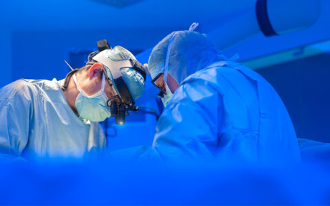
“The most challenging congenital heart defect surgeries are the ones where the surgeon needs to create convoluted alternative pathways for blood flow. VR systems like this can make the procedure clearer, quicker and more reliable,” said David Bichell, M.D., chief of pediatric cardiac surgery at Children’s Hospital.
Patients benefit from less intraoperative decision-making, Bichell added. “If we can determine ahead of time that a certain path isn’t possible, we don’t have to cut into the heart to find that out.”
Bichell recently used Thayer’s system to plan corrective surgery on a patient with complex dextrocardia. “Working from both a 3D printed model of the heart and VR, I understood how it was going to look from behind, how best to move it, how I was going to reach the mitral valve,” he said.
“The most challenging congenital heart defect surgeries are the ones where the surgeon needs to create convoluted alternative pathways for blood flow. VR systems like this can make the procedure clearer, quicker and more reliable.”
Expanding VR Use
To date, only a handful of pediatric cardiac surgery centers routinely use VR, Parra says. The most common application is in training surgical fellows. Parra expects to see expanded adoption for surgical planning as evidence of VR’s benefits accrue and affordable options become more broadly available.
While Bichell’s department is serving as a beta site for his system, Thayer is expanding into other specialties. He is delivering a VR package to Vanderbilt’s radiology team that will enable them to analyze the lungs of patients with pulmonary disease, as well as examine bones. Thayer anticipates embarking on another challenge soon – connecting VR with PET scan imaging for cancer site identification.