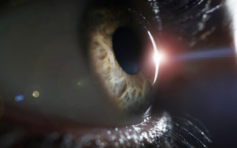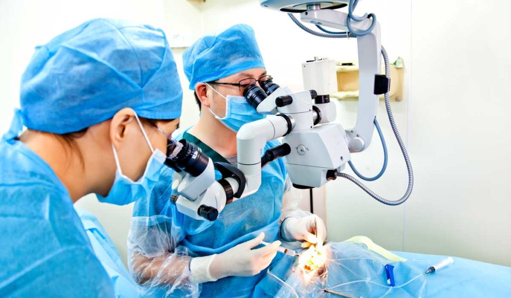
Optical coherence tomography (OCT), a non-invasive imaging technique that provides high-resolution, cross-sectional images, is the gold standard for ophthalmic diagnostics. However, OCT-integrated (iOCT) surgical microscopy technologies that could be used to verify surgical goals have lagged in development due to several fundamental computation and technology limitations.
Under a new five-year $1.8 million grant from the National Eye Institute, a multidisciplinary team of engineers and clinicians at Vanderbilt University Medical Center will advance their work on novel, 4D intraoperative spectrally encoded coherence tomography and reflectometry (iSECTR) technology that allows for simultaneous, intrinsically co-registered and cross-sectional OCT imaging.
“Our group has spent years developing the underlying imaging technology,” said Kenny Tao, Ph.D., an assistant professor of biomedical engineering at Vanderbilt and researcher in the Vanderbilt Institute for Surgery and Engineering (VISE). “This grant will support our translational efforts and allow us to identify ophthalmic surgeries that will directly benefit from real-time guidance.”
“Surgery at advanced stages of eye disease involves precision manipulation of delicate semi-transparent structures in the eye. This advanced imaging could help us better understand how surgical maneuvers impact postoperative visual function and fundamentally change the way we perform ophthalmic surgery,” said Karen Joos, M.D., Joseph and Barbara Ellis Professor of Ophthalmology and Visual Sciences and a VISE researcher.
Advancing iOCT
The Vanderbilt researchers will perform foundational ex vivo and in vivo imaging to quantitatively assess the safety and utility of 4D iSECTR-based surgery. They will also explore novel technologies, feedback mechanisms, and maneuvers to integrate volumetric iSECTR data into image-guided ophthalmic surgery.
“Advances integrating imaging, registration, segmentation and feedback using heads-up display (HUD) visualization will help further address unmet needs in conventional ophthalmic microsurgery.”
“Advances integrating imaging, registration, segmentation and feedback using heads-up display (HUD) visualization will help further address unmet needs in conventional ophthalmic microsurgery,” Tao said.
Clinical Implications
Having comprehensive 4D imaging of tissue-instrument interaction dynamics would provide unprecedented data on structural changes resulting from surgical manipulation. These data could help predict post-operative functional outcomes and enable image-based evaluation of biomechanics and more personalized surgeries. Additionally, iSECTR-based surgery will be compatible with robotic assistance and telemanipulation in the future.
Other clinical collaborators at the Vanderbilt Eye Institute include Shriji Patel, M.D., assistant professor of ophthalmology and visual sciences, and Christine Shieh, M.D., assistant professor of ophthalmology and visual sciences.
A Tool for Public Health
“Eye disorders are expected to increase significantly due to population aging, yet the standard of care to correct visual impairments relies upon intraoperative visualization that hasn’t changed.”
The research has public health implications, says Joos. “Eye disorders are expected to increase significantly due to population aging, yet the standard of care to correct visual impairments relies upon intraoperative visualization that hasn’t changed substantially since our Chairman’s father, Dr. Paul Sternberg, Sr, promoted the use of the surgical microscope for leapfrog improvements in ophthalmic surgeries.”
“Collaboration among engineers and clinicians is what enables the development of [these] practical, clinically useful devices and techniques,” Joos added.