Diagnostic and interventional musculoskeletal ultrasound (MSK US) has ballooned from a niche area of expertise to a recommended treatment modality. The American Medical Society for Sports Medicine found US-guided injections are more accurate, efficacious and, in many cases, more cost-effective than landmark-guided injections for a range of joint and soft tissue conditions.
While the evidence for MSK US is mounting, there is still a shortfall of expert practitioners, says Stephen M. Schaaf, M.D., a physiatrist at Vanderbilt University Medical Center. Schaaf, who joined Vanderbilt in 2019, brings expertise in MSK US to the hospital’s physical medicine and rehabilitation, spine, and sports medicine programs.
“There’s a wide variation in training in musculoskeletal ultrasound, from being capable of performing a knee or shoulder injection to having gone through an accredited fellowship,” Schaaf said. “The gap is most significant in the diagnostic aspect – having the broad knowledge to understand what all musculoskeletal conditions look like normal and abnormal on ultrasound, and how to guide a variety of interventional procedures.”
Advantages and Disadvantages
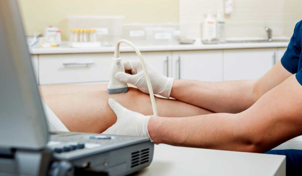
Schaaf says that US can be ideal for diagnosing a wide range of acute and chronic conditions in MSK medicine. This can include joint, bone, tendon, ligament and nerve injuries of the shoulder, elbow, wrist/hand, hip, knee and ankle/foot. The ability to perform point-of-care US as an extension of the patient’s history and physical exam is a key advantage.
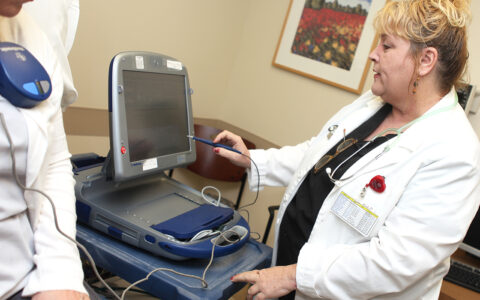
“When a patient comes to my clinic, I have my ultrasound machine right there. I don’t have to send them for an MRI,” Schaaf said. “With the patient present, I can talk with them throughout the examination, show them what I’m scanning, ask what movement hurts that correlates with their symptoms and discuss relevant findings.”
“With the patient present, I can talk with them throughout the examination, show them what I’m scanning, ask what movement hurts that correlates with their symptoms and discuss relevant findings.”
Ultrasound also allows dynamic imaging of MSK structures and the ability to guide injections, Schaaf explains.
“Hip is a common injection that can be done under x-ray. However, I can do it under ultrasound in office while seeing all the neurovasculature around the hip joint,” Schaaf said. “It provides a better understanding of what is a safe approach for that patient, while allowing me to be certain the injection was 100% into the joint.”
Ultrasound is less advantageous when obstructions are present due to the type of injury or the patient’s body habitus. “MRI can still be more effective to diagnose an intraarticular injury, such as pathology inside the knee like an ACL or a meniscus tear,” Schaaf said. “Ultrasound waves can’t penetrate past bone, so it is most effective for superficial soft tissue structures such as tendons, ligaments and nerves around joints.”
Enabling New Approaches
As US technology continues to evolve – offering higher resolution and new imaging modalities – its applications are extending beyond optimizing current diagnostic and interventional approaches to enabling new ones.
Said Schaaf, “With tendon conditions, for example, the research is showing that the pain generator can be micro-invasive nerves and vessels growing into the tendon. With ultrasound guidance, you can perform a percutaneous needle tendon scraping procedure to break up the ingrowth of these micro-nerves and vessels and reset the healing process for that tendon.”
For chronic tendon injuries, another procedure called percutaneous needle tenotomy can be offered as a non-surgical alternative. It involves passing a needle through a tendon with US-guidance to disrupt the degenerative process and promote appropriate tissue healing.
Schaaf also mentions elastography — a new imaging diagnostic modality that uses ultrasound to evaluate the soft tissue characterization of muscle and tendons. “That technology will see increased interest because it gives us the ability to say whether an athlete or a patient is at risk for future injury,” he said.