Most physicians—and even those specializing in diabetes care—likely learned that type 1 diabetes (T1D) is a beta-cell specific autoimmune disorder. Beta cell loss is at the core of the disease with insulin replacement required for survival.
A new study challenges this understanding, by showing that pancreatic volume declines during T1D occurs at a rate that far exceeds beta cell volume.
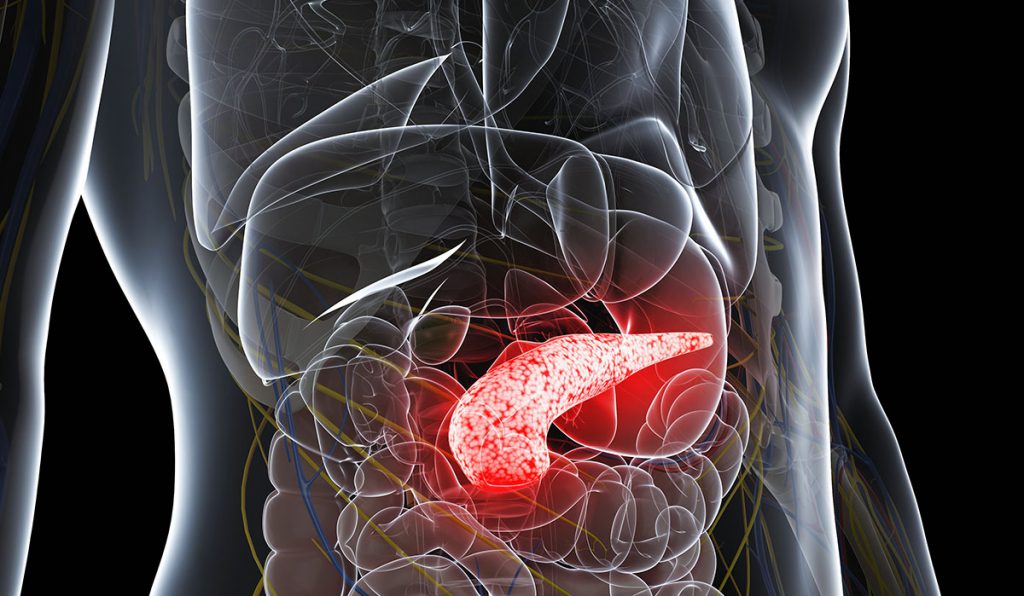
“We think of type 1 diabetes as only the loss of insulin-producing beta cells in the islet. But these cells only account for one to two percent of pancreatic volume. Our study shows pancreatic volume is reduced by up to forty percent,” said Alvin C. Powers, M.D., director of the Vanderbilt Diabetes Center.
Measuring Pancreatic Volume
In results published in Diabetes Care, researchers used MRI to track pancreas volume dynamics in three groups of patients: those with recent-onset stage three T1D, age-matched controls, and autoantibody-positive patients without diabetes. They measured pancreas volume at diagnosis, and six months and one year post-diagnosis. The study involved individuals with diabetes from the Pediatric Diabetes Program in the Eskind Diabetes Clinic and individuals at high-risk for developing type 1 diabetes followed in the Vanderbilt TrialNet Center, headed by William Russell, MD, director of pediatric endocrinology.
In addition to Powers, investigators included former Vanderbilt faculty member Jack Virostko, Ph.D., now at the Dell Medical School Department of Diagnostic Medicine at the University of Texas at Austin; Daniel Moore, M.D., assistant professor of pathology, microbiology and immunology, and pediatrics at Vanderbilt; and Melissa Hilmes, M.D., assistant professor of radiology and pediatrics at Vanderbilt.
A Fundamental Change
“These findings may change how we understand the fundamental biology of T1D.”
Patients with T1D had smaller pancreases than control participants at baseline (median volume 28.6mL versus 48.4mL). Volumes progressively declined in T1D patients over the next year. “These findings may change how we understand the fundamental biology of T1D,” said Moore.
The study suggests other cells must be involved that change the pancreas as a whole, Moore says. This process continues after diagnosis with the volume of the whole pancreas decreasing another 7.2 percent on average over their first year with T1D.
A New Tool to Track Disease
The study could provide a foundation for new diabetes diagnostic and monitoring tools.
“Pancreas size may become an important predictor of T1D onset and could contribute to new ways to understand the response to therapies for T1D, such as by monitoring changes in pancreas volume as a biomarker,” Moore said.
Physicians could use the same non-invasive MRI approach the researchers used in their study to measure treatment responses. It provides an entirely new way to study the role of the whole pancreas in T1D.
The researchers are now working on a multicenter study to test the technology in other health systems. “We’re working with other groups in the US and Australia to standardize the approach to measuring pancreas volume,” Powers said.